As a vet, I happened to do C-Sections to bitches of different breeds.
On these occasions, I noticed some uterus’ defects, directly related to the C-section necessity.
These abnormalities are also called segmental aplasia of Müllerian ducts.
Müllerian ducts are two embryonal organs which develop themselves to form the uterus. Segmental aplasia leads to defects in one uterine horn or in both horns, or in cervix uteri, or vagina’s superior part.
The cases I observed myself can clarify some of these segmental aplasia.
1-Observations
All C-sections have been done at least eight hours after the fall of the progesterone level in blood (checked by laboratory), and after waiting to be sure that the pregnant bitch was unable to whelp by herself.
a-A westie line
-Bitch 1:
First whelping = one pup by C-section.
The pup was located in the left horn which seemed normal.
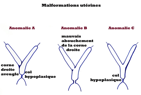
The right horn was empty, without any open exit to the uterus’ body.
The cervix uteri was not normally developed and opened. The internal diameter was not more than 1,5 cm.its walls were thick, without any elasticity, contrarily as it should have been.
Second whelping = one pup by C-section
These abnormalities are called here “Anomalie A”.
-Bitch 2 :
First whelping = one pup by C-section.
The same abnormalities are noticed than the bitch 1.
The left horn contained one pup and was normal.
The right horn was empty and not opened.
The cervix uteri was abnormal, too narrow.
“Anomalie A »
-Bitch 3 :
First whelping = three pups by C-section.
Two pups were located in the left horn, apparently normal.
One pup was located in the right horn, which was not correctly opened into the uterus’ body. The diameter of the horn at the exit was about 1,5 cm. this pup could not go through without a c-section.
The cervix uteri was normal
These abnormalities are called here “Anomalie B”.
Second whelping = three pups by C-section
Two pups in the left horn.
One pup in the right horn
b-Other westie lines
First case = bitch with one pup by C-section. “ Anomalie A”
Second case = bitch with three ,pups by C-section.
Normal horns.
Cervix uteri with small diameter and thick walls without any elasticity.
This abnormality is called here “Anomalie C”.
c-Other breed
Clear Cream Labrador bitch = one pup by C-section = “Anomalie A”
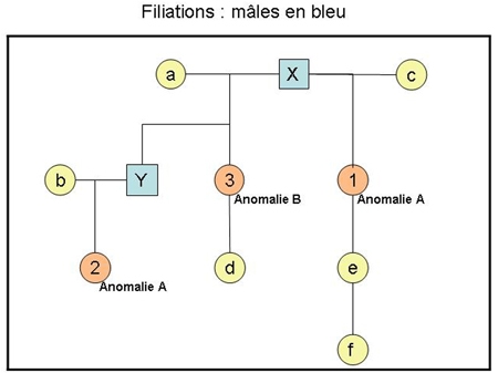
2- Family connections, between bitches 1,2 and 3 from the same line :
3- Formation and malformation of uterus
During embryogenesis, the two Müllerian ducts grow in the direction of the urogenital sinus and also of one each other. They cling together and to the sinus to form the uterus’ horns, the uterus’ body, the cervix uteri, and part of vagina, when walls are resorbed.
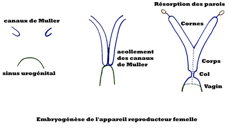
Abnormalities A,B and C occur during this process :
.
4- « White Heiffer Disease »,
This hereditary disease is a congenital atresia which can concern oviducts, uterus or vagina, due to a segmental aplasia of Müllerian ducts.
This was described principally in “Blanc Bleu Belge” and “british Shorthorn” breeds. It had been well studied and a recessive gene has been identified, which can express itself when the “white shorthorn factor” is present.
The recessive gene of this disease is called mdh01.
The cows with this double recessive gene, “homozygote recessives”, have vaginal agenesis or more rarely uterine agenesis.
Uterine agenesis are segmental aplasia more or less pronounced, concerning both horns or just one horn (more frequently the right one), or cervix uteri more rarely.
This disease appears only if the cow is white coloured. This colour is due to another recessive gene, called White Shorthorn Factor. The locus name is R, for Roan colour. This gene is called kitlg, or mgf. The homozygote cows with the dominant alleles are uniform colours (red for shorthorn breed, and grey for the BBB breed).
The heterozygote cows wear a mixing of white and coloured hairs (Roan colour for the Shorthorn, and blue colour for the BBB).
Only the white cows, recessive homozygote for two genes, (mgf and mdh01), can show this disease, which concerns a few number of the white cows.
5-Conclusion for dogs.
The abnormalities observed in
dogs show similarities to the cows, even though the uterus is more
likely to be concerned for dogs whereas vagina is more likely to be
concerned for cows.
In the case of the uterus, the
right horn is more likely to be concerned for both species. This leads to consider that an
autosomal recessive gene could be involved for dogs, as for cows.
A recessive colour gene could be
also responsible, as for cows, for the appearance of the defect, as a
favouring factor. At this point, all the cases I observed were white or
cream dogs (very clear cream Labrador, almost white).
Perhaps special studies in
Labradors could help to conclude, and also in other terrier breeds, like
Scotties and Cairns, cousins to Westies, and showing different colours.
Dr Patricia Picquot.